Pterygopalatine fossa 7
Share your inquiries now with community members
Click Here
Sign up Now
Lessons List | 29
Lesson
Comments
Our New Certified Courses Will Reach You in Our Telegram Channel
Join Our Telegram Channels to Get Best Free Courses
Join Now
We Appreciate Your Feedback
1 Reviews

Belyu mekonen
Show More Reviews
Related Courses in Medical

Course Description
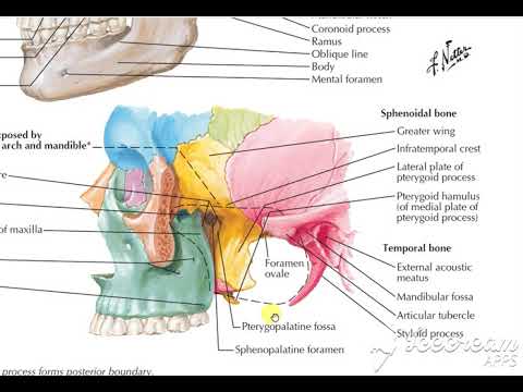
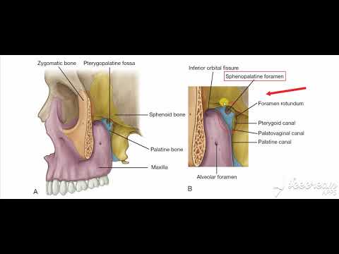
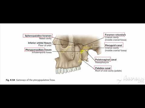
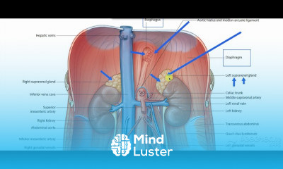
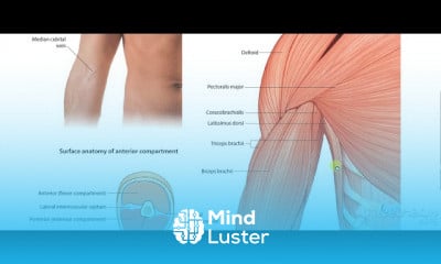
In human anatomy, the pterygopalatine fossa (sphenopalatine fossa) is a fossa in the skull. A human skull contains two pterygopalatine fossae—one on the left side, and another on the right side. Each fossa is a cone-shaped paired depression deep to the infratemporal fossa and posterior to the maxilla on each side of the skull, located between the pterygoid process and the maxillary tuberosity close to the apex of the orbit. It is the indented area medial to the pterygomaxillary fissure leading into the sphenopalatine foramen. It communicates with the nasal and oral cavities, infratemporal fossa, orbit, pharynx, and middle cranial fossa through eight foramina.
The pterygopalatine fossa is a bilateral, cone-shaped depression extending deep from the infratemporal fossa all the way to the nasal cavity via the sphenopalatine foramen.
It is located between the maxilla, sphenoid and palatine bones, and communicates with other regions of the skull and facial skeleton via several canals and foramina. Its small volume combined with the numerous structures that pass through makes this a complex region for anatomy students.
This article will discuss the pterygopalatine fossa, and consider the structures involved according to their respective foramina.
By TeachMeSeries Ltd (2021)
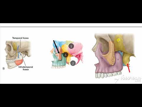
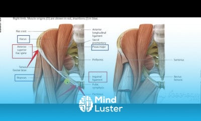
Fig 1.0 - Left infratemoporal fossa demonstrating the opening of the pterygopalatine fossa (circled in red). Note: the zygomatic arch has been removed in this image.
Fig 1.0 – Left infratemoporal fossa demonstrating the opening of the pterygopalatine fossa (circled in red). Note: the zygomatic arch has been removed in this image.
Borders
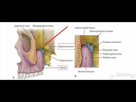
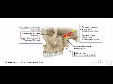
The borders of the pterygopalatine fossa are formed by the palatine, maxilla and sphenoid bones:
Anterior: Posterior wall of the maxillary sinus.
Posterior: Pterygoid process of the sphenoid bone.
Inferior: Palatine bone and palatine canals.
Superior: Inferior orbital fissure of the eye.
Medial: Perpendicular plate of the palatine bone
Lateral: Pterygomaxillary fissure
Contents
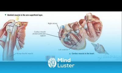
The Pterygopalatine Fossa contains many important neurovascular structures. Here we will discuss the maxillary nerve and its branches, the pterygopalatine ganglion and the maxillary artery and its branches.
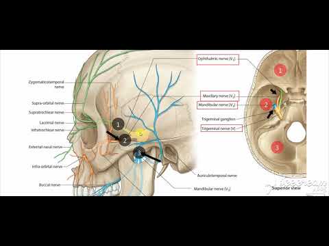
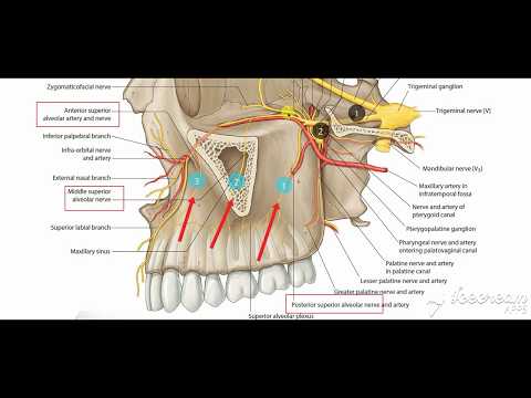
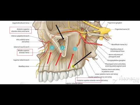
Maxillary Nerve
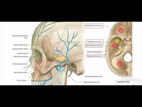
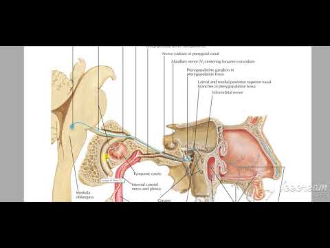
The maxillary nerve is the second branch of the trigeminal nerve (CNV2). It passes from the middle cranial fossa into the pterygopalatine fossa through the foramen rotundum.
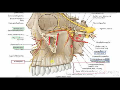
The main trunk of the maxillary nerve leaves the pterygopalatine fossa via the infraorbital fissure. Here, it enters the infraorbital canal of the maxilla and exits below the orbit in the infraorbital foramen to contribute to the sensory innervation of the face (figures 2.0 & 2.1).
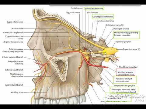
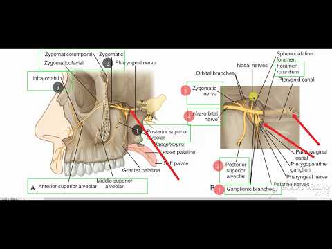
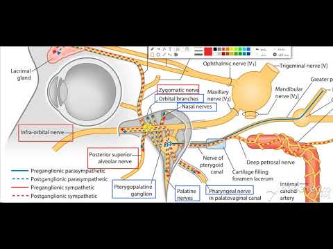
While in the pterygopalatine fossa, the maxillary nerve gives of numerous branches including the infraorbital, zygomatic, nasopalatine, superior alveolar, pharyngeal and the greater and lesser palatine nerves. The maxillary nerve also communicates with the pterygopalatine ganglion (discussed below) via two small trunks, the pterygopalatine nerves (figure 2.1). These nerves suspend the ganglion within the pterygopalatine fossa.
By TeachMeSeries Ltd (2021)
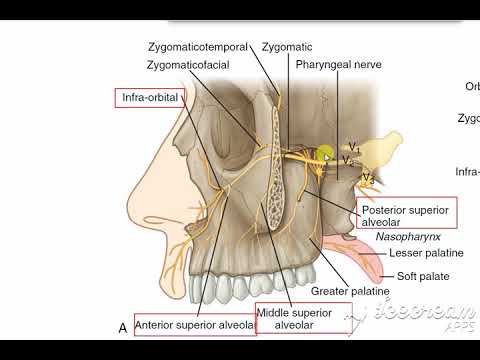
Fig 3.0 - The main trunk of the maxillary nerve (CNV2); showing the origin at the trigeminal nerve and its path to external facial structures.
Fig 2.0 – The main trunk of the maxillary nerve (CNV2); showing the origin at the trigeminal nerve and its path to external facial structures.
By TeachMeSeries Ltd (2021)
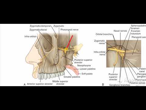
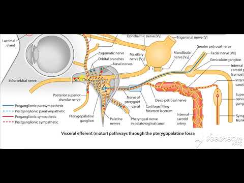
Fig 3.1 - The branches of the pterygopalatine ganglion and the maxillary nerve. Note: For simplicity, this schematic does not show: the contribution of the facial nerve (CNVII) to the pterygopalatine ganglion, the posterior superior alveolar nerves, or the nerve of the pterygoid canal.
Fig 2.1 – The branches of the pterygopalatine ganglion and the maxillary nerve. Note: For simplicity, this schematic does not show: the contribution of the facial nerve (CNVII) to the pterygopalatine ganglion, the posterior superior alveolar nerves, or the nerve of the pterygoid canal.
Pterygopalatine Ganglion
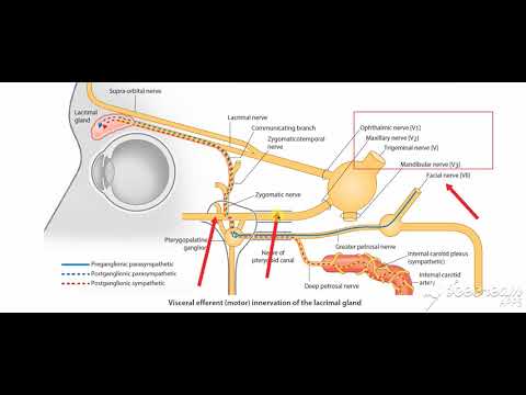
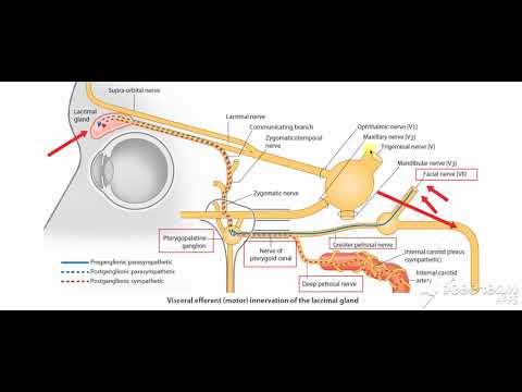
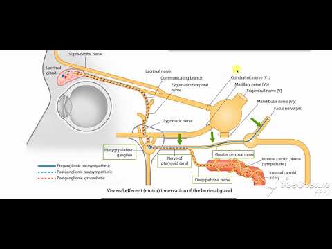
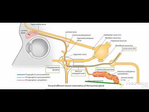
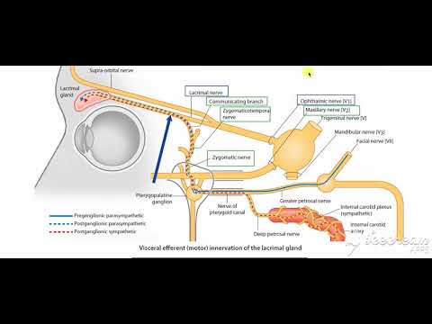
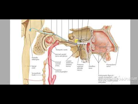
The pterygopalatine ganglion sits deep within the pterygopalatine fossa near the sphenopalatine foramen. It is the largest parasympathetic ganglion related to branches of the maxillary nerve (via pterygopalatine branches) and is predominantly innervated by the greater petrosal branch of the facial nerve (CNVII).
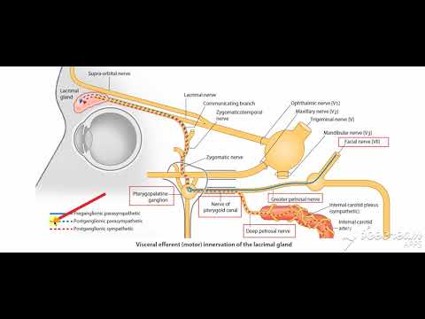
Postsynaptic parasympathetic fibres leave the ganglion and distribute with branches of the maxillary nerve (CNV2). These fibres are secretomotor in function, and provide parasympathetic innervation to the lacrimal gland, and muscosal glands of the oral cavity, nose and pharynx.
By TeachMeSeries Ltd (2021)
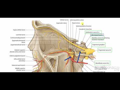
Fig 1.2 - The pterygopalatine ganglion and its branches.
Fig 3.0 – The pterygopalatine ganglion and its branches.
Maxillary Artery
The maxillary artery is a terminal branch of the external carotid artery. The terminal portion of the maxillary artery lies within the pterygopalatine fossa. Here, it separates into several branches which travel through other openings within the fossa to reach the regions they supply.
These branches include, but are not limited to:
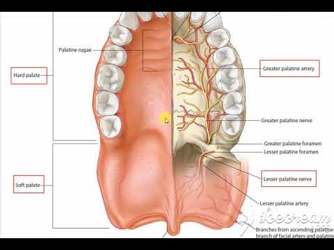
Sphenopalatine artery (to the nasal cavity).
Descending palatine artery – branches into greater and lesser palatine arteries (hard and soft palates).
Infraorbital artery (lacrimal gland, and some muscles of the eye).
Posterior superior alveolar artery (to the teeth and gingiva).
At their terminal ends, the sphenopalatine and greater palatine arteries anastomose at the nasal septum.
Trends
WiFi hacking
MS Excel
C Programming Language
Adobe illustrator tools for designers
Ethical Hacking
Python programming language
Digital Electronics basics
Excel Course Basic to Advanced
Learning English Speaking
Mobile Apps from Scratch
AI Writing tools in google docs for beginners
Digital Marketing
Complete WIFI Hacking Course Beginner to Advanced
Excel Power Query in excel for beginners
Human Resource Management
Generative AI Tutorial For Beginners
Graphic design rules for beginners
Infection Control
The Complete Python Programming Full Course
Accounting Finance course
Recent
Rock sitting dance workout for beginners
Fat Burning home workout For beginners
Home weight loss workout For women
Cardio Kickboxing HIIT for beginners
HIIT cardio walking workout at home
Gospel cardio dance workout at home
Upper body cardio weights for beginners
Legs workout with dumbbell for beginners
Cardio workout for beginners
Hotel HIIT legs workout for beginners
Cool down stretch workouts at home
Cardio boxing workout at gym
Legs and abs workout at home
Cardio Kickboxing workouts at gym
Pilates and barre inspired workouts at gym
Intense cardio HIIT workouts at gym
Knockout cardio workouts for beginners
Tucked abs workout at home
Hamstrings tempo workout at home
Dumbbell cardio HIIT workouts at gym