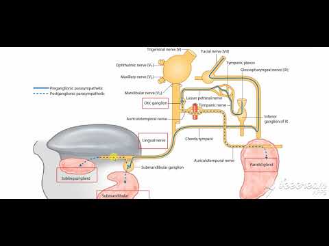
Submandibular salivary gland nerve supply 1
Share your inquiries now with community members
Click Here
Sign up Now
Lessons List | 14
Lesson
Comments
Our New Certified Courses Will Reach You in Our Telegram Channel
Join Our Telegram Channels to Get Best Free Courses
Join Now
We Appreciate Your Feedback
11 Reviews

KUSHAGRA PANDEY

Bhanu pal

Dr. M. K. Goel

AVIK MANDAL
Show More Reviews
Related Courses in Medical
Course Description
The submandibular gland is the second largest of the three main salivary glands, which also include the parotid and sublingual glands. The submandibular glands are paired major salivary glands that lie in the submandibular triangle. The glands have a superficial and deep lobe separated by the mylohyoid muscle
The Wharton duct, the submandibular gland’s primary excretory duct, drains into the oral cavity at the sublingual caruncle. The sublingual caruncle is a papilla located medial to the sublingual gland and lateral to each side of the frenulum linguae [1]. The submandibular gland produces approximately 70% of the saliva in the unstimulated state. However, the parotid gland’s saliva production predominates once the salivary glands become stimulated
The paired submandibular glands (historically known as submaxillary glands) are major salivary glands located beneath the floor of the mouth. They each weigh about 15 grams and contribute some 60–67% of unstimulated saliva secretion; on stimulation their contribution decreases in proportion as the parotid secretion rises to 50%. The average length of the normal human submandibular salivary gland is approximately 27mm, while the average width is approximately 14.3mm.
The submandibular glands are bilateral salivary glands located in the face.
Their mixed serous and mucous salivary secretions are important for the lubrication of food during mastication to enable effective swallowing and aid digestion.
In this article, we shall look at the anatomy of the submandibular gland – its location, blood supply and clinical correlations.
The submandibular gland is located within the anterior part of the submandibular triangle. The boundaries of this triangle are:
Superiorly: Inferior body of the mandible.
Anteriorly: Anterior belly of the digastric muscle.
Posteriorly: Posterior belly of the digastric muscle.
Anatomical Structure
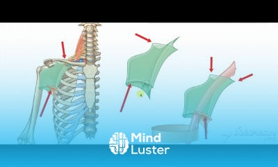
Structurally, the submandibular glands are a pair of elongate, flattened hooks which have two sets of arms; superficial and deep. The positioning of these arms is in relation to the mylohyoid muscle, which the gland hooks around.
Superficial arm – comprises the greater portion of the gland and lies partially inferior to the posterior half of the mandible, within an impression on its medial aspect (the submandibular fossa). It is situated outside the boundaries of the oral cavity.
Deep arm – hooks around the posterior margin of mylohyoid through a triangular aperture to enter the oral cavity proper. It lies on the lateral surface of the hyoglossus, lateral to the root of the tongue.
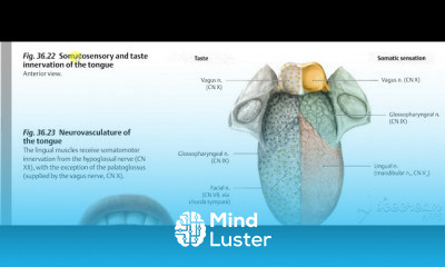
Secretions from the submandibular glands travel into the oral cavity via the submandibular duct (Wharton’s duct). This is approximately 5cm in length and emerges anteromedially from the deep arm of the gland between the mylohyoid, hypoglossus and genioglossus muscles. The duct ascends on its course to open as 1-3 orifices on a small sublingual papilla (caruncle) at the base of the lingual frenulum bilaterally.
Trends
Web design basics
Cybersecurity fundamentals A Z
Accounting Finance course
E Commerce web design
Web Design for Beginners
Create Animals icon in figma
UX UI design career
Customizing type for logos
Essential skills for web designers
UX design fundamentals
Create food and drink icon in figma
Figma web design
Best zoology books
Create a YouTube account on Your phone
Figma mobile UI design essentials
Figma mobile app design
SQL for accountants and finance managers
Web Design 101 Free Full Course
Abstract Poster design in figma
Mastering logo design in illustrator
Recent
Bioinformatics basics
Bioinformatics databases
Vitamin A to Z tablets
Best zoology books
Best cream for piles pain
Laser surgery for piles
Best cream for piles
Anal fissure treatment
Best antibiotics for diseases
Antibodies structure
Macrophage structure
Drosophila genetics
Diagnostic tests
Bioinformatics
Genetics
Gene therapy
Kidney structure
DNA replication and types
Bacterial cell structure
Parasite structure